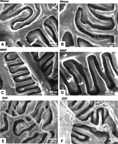
Figure 6.
The ultrastructure of the filtration slits is similar in different rat strains, both in normal and proteinuric conditions. Representative scanning electron photomicrographs of filtration slit ultrastructure between neighboring podocytes in Wistar (A and B), MWF (C and D), and ZDF (E and F) rats, taken with an in-lens detector. Original magnifications: A and C, 120,000×; B, 85,000×; D, 150,000×; E, 100,000×; F, 140,000×.

