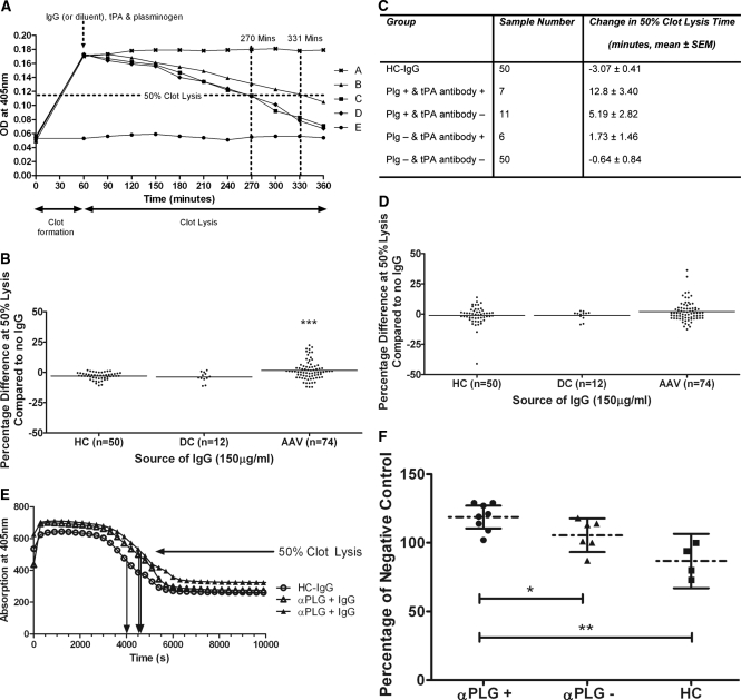
Figure 4.
Anti-plasminogen antibodies in AAV patient IgG compromise fibrinolysis. (A) Fibrin clot formation was stimulated by mixing thrombin with fibrinogen and monitored spectrophotometrically. No clot was formed in the absence of thrombin (●). Sample diluent (■), healthy control IgG (♦), or UK AAV-IgG (▴) were incubated with plasminogen (PLG) before the addition of tissue plasminogen activator (tPA). The tPA-containing solution was immediately loaded onto the fibrin clot. Fibrinolysis was measured by monitoring the reduction in absorbance at 405 nm. Plasminogen alone did not initiate fibrinolysis (x), whereas AAV-IgG retarded fibrinolysis compared with sample diluent and control IgG (▴). (B) Time to 50% fibrinolysis was retarded by 18/74 (24.3%) UK patient IgG compared with none of the healthy or disease controls. ***P < 0.001 AAV-IgG compared with healthy control IgG. (C) Average change in 50% clot lysis times (UK patients). (D) When IgG was premixed with plasmin (PLM) on ice before loading onto a fibrin clot, 5/74 (6.7%) UK AAV-IgG retarded fibrinolysis (2 from MPO-ANCA and 3 from PR3-ANCA patients). (E) In separate experiments, Dutch AAV-IgG samples were preincubated with PLG and mixed with fibrinogen, factor XIII, and tPA. This mixture was subsequently added to thrombin and CaCl2. Fibrin clot formation and lysis, monitored by change in absorbance at 405 nm, are shown for two anti-plasminogen antibody–positive AAV-IgG and one healthy control. Arrows indicate 50% lysis times. (F) Half-lysis times are given as percentage of a negative control with AAV-IgG grouped by anti-plasminogen antibody status (mean and 95% confidence intervals).

