Figure 9.
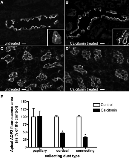
Indirect immunofluorescence of sections from rat kidney showing AQP2 in principal cells of cortical and inner medullary collecting ducts with and without CT treatment in vivo. After implantation of minipumps containing either saline or CT for 4 hours, the animals were anesthetized, and the kidneys were fixed by perfusion, followed by sectioning and immunostaining to detect AQP2. (A through C) Under control conditions, AQP2 is localized toward the apical pole of collecting ducts from the cortex (A) and inner medulla (C), as well as the cortical connecting segment (A, inset), but in the presence of CT, AQP2 is more sharply concentrated at the apical pole of principal cells from the cortical collecting duct (B) and in cells of the connecting segment (B, inset), reflecting a reduction in cytoplasmic vesicle staining and an increase in apical membrane staining (see also Figure 6). (D) CT had no effect in the inner medulla, where AQP2 remained localized throughout the cytoplasm. (E) Quantification of the effect of CT on AQP2 redistribution in principal cells showed a significant increase in apical staining in both cortical collecting ducts and, to an even greater extent, connecting segments. No effect was detected in the inner medulla. The images are representative of three independent incubations, and quantification shows the mean of more then 100 cells taken from the three different experiments (means ± SEM; *P < 0.05). The bar indicates 10 μm.

