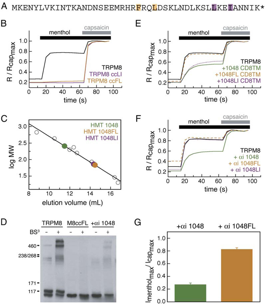
Figure 6. Mutations in TRPM8 Coiled-Coil Core Alter Oligomerization and Channel Function.
(A) TRPM8 coiled-coil sequence. Positions of “a” and “d” mutations are indicated, FL→AA (orange) and LI→AA (purple).
(B) Normalized calcium imaging traces from 303 to 494 capsaicin-sensitive cells expressing TRPM8, TRPM8 and TRPM8-ccFL, and TRPM8 and TRPM8-ccLI.
(C) Gel filtration of FL and LI mutant HMT fusion proteins. Wild-type coiled coil (HMT-1048) is shown in green for comparison. HMT-1048FL (orange) and HMT-1048LI (purple) elute as monomers.
(D) BS3 crosslinking of wild-type TRPM8, full-length TRPM8 bearing the FL coiled-coil mutation (TRPM8ccFL), and TRPM8 coexpressed with αi 1048. No specific crosslinking is seen with TRPM8ccFL. Coexpression of αi 1048 does not prohibit crosslinking of TRPM8.
(E) Normalized calcium imaging traces from HEK293T cells cotransfected with CD8 fusions: 1048 CD8 (green), 1048LI CD8TM (purple), and 1048FL CD8TM (orange). Each trace is an average of 376 to 536 cells.
(F) Normalized calcium imaging traces cells cotransfected with αi fusions, αi-1048 (green), αi-1048FL (orange), and αi-1048LI (purple). Each trace is an average of 398 to 830 cells.
(G) Average maximal menthol responses normalized to maximal capsaicin responses for oocytes expressing TRPM8, TRPV1, and the indicated αi fusions. Standard errors are indicated (p < 0.001, n = 9–12 cells).

