Figure 5.
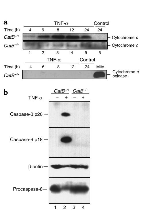
(a) TNF-α–induced release of cytochrome c into the cytosol is reduced in catB–/– mouse hepatocytes. At the indicated times after addition of medium lacking (control) or containing TNF-α + AcD, cytosol fractions were prepared by selective permeabilization with digitonin as described in Methods. Aliquots containing 20 μg of protein were subjected to SDS-PAGE on gels containing 15% acrylamide, transferred to nitrocellulose, and probed for cytochrome c. Samples from catB+/+ hepatocytes were also probed for cytochrome c oxidase, to exclude a possible mitochondrial contamination in the cytosol. (b) Caspase-9 and caspase-3 are processed in TNF-α/AcD–treated catB+/+ hepatocytes but not in catB–/– hepatocytes. Cells were incubated for 24 hours in medium lacking or containing TNF-α + AcD. After whole-cell lysates were prepared as described in Methods, aliquots containing 50 μg protein were subjected to SDS-PAGE, transferred to nitrocellulose membrane, and analyzed by immunoblot using antisera that recognize only active caspase-9 or active caspase-3 (36). The same blot was probed with sera that recognize procaspase-8 and β-actin to confirm loading and transfer of samples from catB–/– mice.

