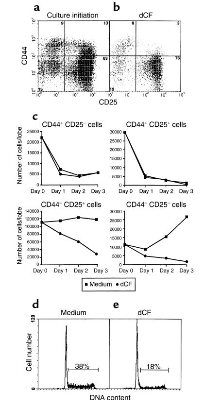
Figure 2.
Characterization of thymocytes from ADA-inhibited FTOCs. CD25 and CD44 expression were compared in freshly isolated thymocytes from C57BL/6 fetuses at day 15 of gestation (a) and in those harvested from day 15 FTOCs after 5 days of culture with 5 μM dCF (b). Single-cell suspensions were stained with Cy-Chrome-rat anti-CD4, Cy-Chrome-rat anti-CD8, and FITC-rat anti-CD25 followed by PE-rat anti-CD44. Finally, the stained cells were incubated with PI to allow the exclusion of dead cells. CD25 versus CD44 (a and b) staining of cells negative for PI and Cy-chrome staining is shown for one of three representative experiments. In other experiments (c), cells were harvested on days 1, 2, and 3 of culture, counted, and then stained with PE-anti-CD4, PE-anti-CD8, PE-anti-γδ TCR, FITC-anti-CD25, and biotinylated anti-CD44 plus RED 613-streptavidin. Cells expressing CD4, CD8, and/or γδ TCR (i.e., PE-labeled cells) were excluded from the analysis, and CD44 and CD25 were assessed on the remaining double negative population. In other experiments, thymocytes were harvested from day 15 FTOCs after 2 days of culture in the presence (e) or absence (d) of 5 μM dCF. Cell-cycle analysis was performed by PI staining, and DNA content was evaluated with a FACScan. Data from one representative experiment of four are shown.

