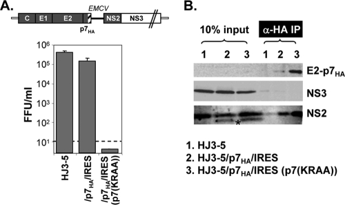
FIG. 5.
Infectious virus release from HJ3-5/p7HA-IRES-transfected cells and p7HA-mediated pulldown of NS2 and NS3 proteins. (A) The upper panel shows the organization of HJ3-5/p7HA-IRES. The lower panel shows infectious virus release into cell culture fluid at day 2 postelectroporation of the indicated RNA into FT3-7 cells. Error bars indicate standard deviations. (B) Western analysis of proteins following anti-HA IP. The proteins were run on 10% PAGE and probed with anti-HA, NS3, or NS2 antibodies, as noted. The asterisk indicates an additional band detectable by NS2 antibody. We speculate that this band may represent an aberrant initiation product by EMCV IRES, since this band appeared only from HJ3-5/p7HA/IRES-related constructs.

