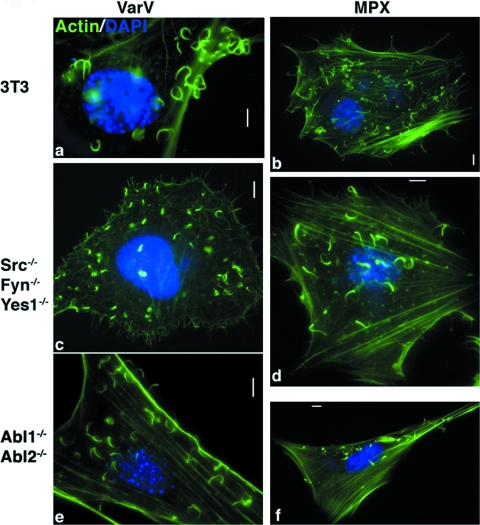
FIG. 2.
VarV and MPX form actin tails. (a to f) Images of fibroblast cell lines derived from WT mice (3T3) (a and b), Src−/− Fyn−/− Yes1−/− mice (c and d), and Abl1−/− Abl2−/− mice (e and f), infected with VarV strain BSH (a, c, and e) or with MPX (b, d, and f) for 24 h, fixed, and stained with DAPI (blue) and FITC-phalloidin (green) to recognize DNA and actin, respectively. Bars, 5 μm.

