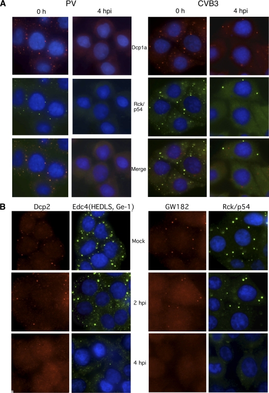
FIG. 1.
Disruption of cytoplasmic P bodies in HeLa cells during PV infection. (A) HeLa cells were infected with PV (left) or CVB3 (right) at an MOI of 20 for 4 h before fixation and processing for immunofluorescence by staining with antibodies against Rck/p54 and Dcp1a as markers for P bodies. (B) HeLa cells infected with PV for various times were processed as described for panel A before dual staining with antibodies to P body marker proteins Dcp2 and Edc4 or GW182 and Rck/p54 as indicated.

