FIG. 8.
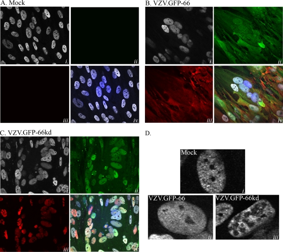
The ORF66 kinase influences matrin 3 nuclear distribution in VZV infections. Shown are immunofluorescence analyses of mock-infected MRC-5 cells (A) and of MRC-5 cells infected with VZV.GFP-66 (B) or VZV.GFP-66kd (C) at an MOI of 0.003. Cells were fixed with 4% paraformaldehyde 3 days postinfection and were immunostained either with a rabbit α-matrin 3 antibody (A to C, panels i, and D), detected with α-rabbit Alexa Fluor 546, or with mouse α-IE62 (A to C, panels iii), detected with α-mouse Alexa Fluor 647. ORF66 expression was determined by GFP autofluorescence (A to C, panels ii). The merge images (A to C, panels iv) are overlays of matrin 3 (gray), GFP (green), IE62 (red), and nuclei stained with Hoechst dye (blue). (D) Representative single-cell images depicting matrin 3 localization. Infection conditions are given on the images. Fluorescence images were taken using a 60× objective.

