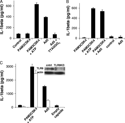
FIG. 3.
TLR9 recognition of Ad5 DNA contributes to IL-1β release from human but not mouse macrophages. (A) hMDMs were treated with control medium, primed with PAM3CSK4 for 4 h before medium with or without ATP was replaced for 1 h, or treated with Ad5gfp in the presence or absence of 20 μM of the TLR9 inhibitor (TTAGGG)4 for 8 h. (B) mBMMs were either primed with PAM3CSK4 for 4 h before being treated with control medium (4 h), ATP (1 h), or Ad5gfp (4 h) or left unprimed and treated with control medium or Ad5gfp for 8 h. (C) PMA-differentiated THP-1cntrl (closed) or THP-1tlr9KD (open) cells were treated with control medium for 6 h, primed with PAM3CSK4 before treatment with ATP for 1 h, or treated with Ad5gfp alone or an equivalent amount of empty capsids for 6 h, as described in Materials and Methods. The release of IL-1β was quantified by ELISA. Data represent the means and standard errors from 3 replicates. (Inset) Western blot for TLR9 and the actin loading control in THP-1cntrl and THP-1tlr9KD cell lysates.

