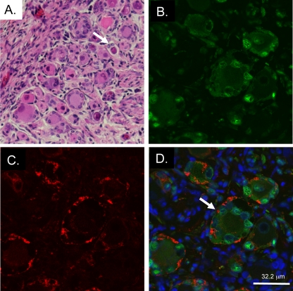
FIG. 9.
Histopathological effects of DRG xenograft infection with gE-AYRV. (A) gE-AYRV-infected xenograft at 14 days postinfection, with H&E staining (magnification, ×200). The white arrow indicates a cytopathic effect in the neuron, i.e., contracted cytoplasm and inclusion bodies. (B to D) gE-AYRV-infected xenograft stained with rabbit polyclonal antibody to IE63 (B) and mouse monoclonal antibody to gE (C) and merged image with Hoechst counterstain (D). The white arrow in D indicates the patchy appearance of membrane-associated gE.

