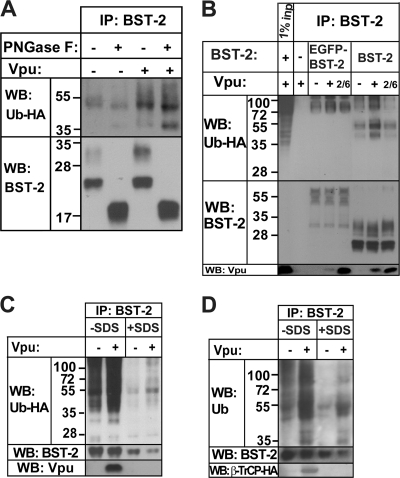
FIG. 2.
The ubiquitinated species in BST-2 immunoprecipitates are likely BST-2 itself. (A) The transfection of 293T cells and IP was performed as described in the legend of Fig. 1A. Sodium phosphate (to a concentration of 50 mM) and NP-40 (to a concentration of 1%) were added directly to the IPs in Laemmli buffer before treatment with PNGase F. For each 10-cm2 dish, 2.5 units of PNGase F (Sigma) was added, followed by incubation at 37°C for 6 h. (B) The transfection of 293T cells and IP were performed as described in the legend of Fig. 1A, except that EGFP-BST-2 was used. Cells transfected with empty EGFP plasmid were used as a no-BST-2 negative control. (C) The transfection of 293T cells and IP were performed as described in the legend of Fig. 1A. IPs were eluted in Laemmli buffer, with boiling. Half of the sample was diluted 35 times with lysis buffer and reimmunoprecipitated using monoclonal anti-BST-2 antibody prebound to anti-mouse IgG magnetic beads. (D) The transfection of 293T cells was performed as described in the legend of Fig. 1B, and IP was performed as described in the legend of Fig. 2C, except that two-thirds of the sample was used for reimmunoprecipitation.

