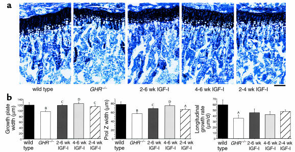
Figure 5.
Chondrocyte proliferation is restored in GHR–/– mice treated with IGF-I. (a) Representative proximal tibial sections stained with toluidine blue from 6-week-old wild-type, GHR–/–, and GHR–/– mice treated with IGF-I from weeks 2–6, 4–6, and 2–4. Scale bar, 100 μm. (b) Tibial growth-plate width, proliferative-zone width, and longitudinal growth rate were increased in IGF-I–treated mice. n = 4–10 per group. AP < 0.01, BP < 0.001 vs. wild-type, CP < 0.05, DP < 0.01 vs. GHR–/–.

