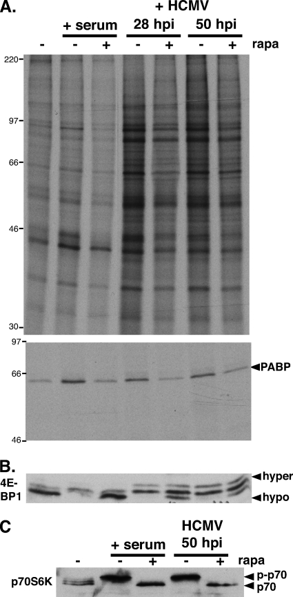
FIG. 4.
Rapamycin sensitivity of HCMV-induced PABP mRNA translation and p70RSK activation. (A, top) Growth-arrested NHDFs were either serum stimulated (+ serum) or infected with HCMV (MOI = 5). At 26 or 46 hpi, HCMV-infected cells were treated with either DMSO or rapamycin (rapa) for 30 min and subsequently pulse-labeled with [35S]Met-Cys with (+) or without (−) rapa for 1.5 h. Uninfected cells (with or without rapa) were similarly metabolically labeled following stimulation with 20% FBS for 20 min. Total protein (top) and PABP immunoprecipitates (bottom) were fractionated by SDS-PAGE and visualized by autoradiography. The migration of molecular mass standards (in kilodaltons) is shown on the left. (B, C) Lysates from panel A were fractionated by SDS-PAGE and analyzed by immunoblotting using the indicated antibodies. The hyper- and hypophosphorylated 4E-BP1 forms are designated. p-p70, phosphorylated p70 S6K.

