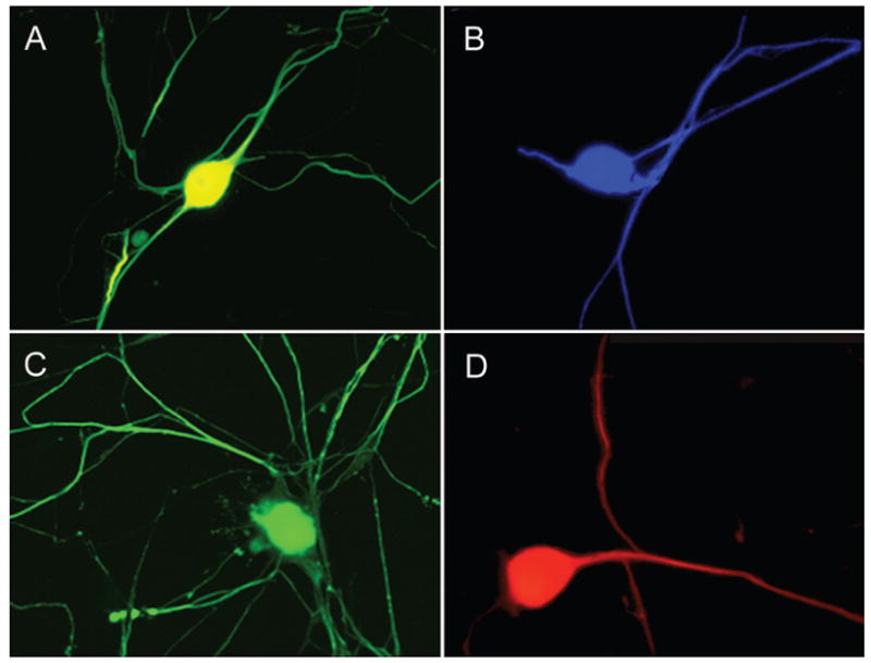
Fig. 2.

Representative fluorescence micrograph of the center region of a 1-cm long living nervous tissue construct engineered from harvested adult human DRG neurons. To create this construct, 2 populations of the neurons were plated on adjacent membranes in an axon elongation apparatus. Axons grew out across the ~ 100-μm gap between the 2 membranes to integrate with the neuron population on the other side. Subsequently, continuous tension was placed on the bridging axons at an escalating rate, by mechanically separating the 2 membranes. This process induced axon “stretch-growth” of up to 1 mm/day and promoted fasciculation of the axons into large bundles reaching 1 cm in length. The stretch-grown axon bundles are demonstrated by immunoreactivity to an antibody specific for phosphorylated neurofilament protein (SMI-31).
