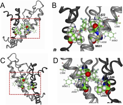
Fig. 7.
NS1643 docked to a homology model of two adjacent subunits of a hERG1 channel. A, side view of two adjacent hERG1 subunits highlighting the residues that were identified as important for drug activity based on the results of the voltage-clamp assay for hERG1 mutations as summarized in Fig. 2. NS1643 is portrayed in thick ball-and-stick mode and is located in the middle of the region delineated by the dashed box. The pore helix, selectivity filter, and S6 segment of one subunit is colored dark gray; the S4–S5 linker, S5, pore helix, selectivity filter, and S6 of an adjacent residue is colored light gray. The turret region (between pore helix and S6 segment) was not modeled and represents the structure of the Kv1.2 channel. B, amplified view of the region inside the boxed region shown in A. Residues identified by scanning mutagenesis as the most important for interaction with NS1643 are labeled. Boldface text was used for the residues shown in space-fill that are in close proximity to the drug. Smaller font and italicized text identify residues (thin ball-and-stick mode) that are not in close proximity to the drug. C and D, similar to A and B, but after 180° rotation.

