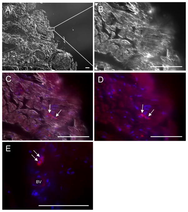
Figure 3.
The survival of MSCs in the penile sections using differential interference contrast (DIC) to define tissue morphology and epifluorescence to localize GFP. A, DIC image from edge of penis section. B, DIC image from edge of penis section. C, DIC image merged with staining by anti-GFP antibody (ALEXA 594, red, TRITC channel) and DAPI to visualize cell nuclei (blue). D, Epifluoresence alone from image in (C) showing signals for GFP and DAPI. E, Epifluoresence image from a different p75dMSC-injected rat in which the engrafted cells were located in close proximity to a blood vessel. BV: Blood vessel. Scale bars= 100 micrometers.

