
Figure 4.
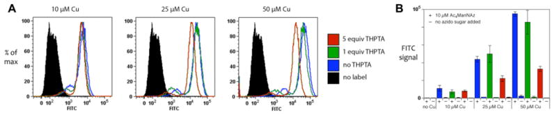
Flow cytometry analysis of Jurkat cells, pre-incubated for 48 h in growth medium containing 10 μM Ac4ManNAz, and then labeled with 3 (50 μM) under standard conditions (CuSO4 and THPTA at the indicated concentrations, 2.5 mM Na ascorbate, 1 mM aminoguanidine, in DPBS, 4 °C, 5 min). (A) All samples were analyzed in triplicate; 10,000 events each were recorded in each case, and representative histograms are shown. (B) Mean fluorescence intensities with error bars representing standard deviations (statistical analysis performed using FlowJo software 8.7.1).