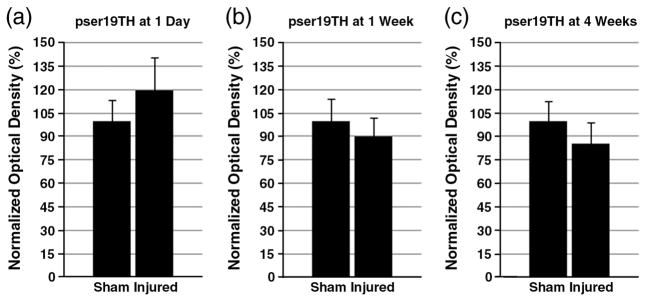
Fig. 1.
Western blot of striatal pser19TH. The levels of striatal pser19TH after CCI or sham injury were analyzed by Western blotting. Optical densities of each phosphorylated protein were normalized by β-actin, and each group had n=6. The levels of TH at 1 day (a), 1 week (b), and 4 weeks after injury are summarized here. Each normalized optical density±SEM is displayed. There was no alteration of pser19TH at any of the time points.

