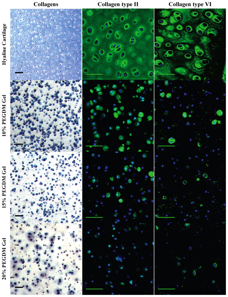
Figure 3.
Gross examination of collagen matrix deposition by histological and immunohistochemical (IHC) evaluation for chondrocytes encapsulated in 10, 15, or 20% PEG gels and cultured for 4 weeks. Sections were stained for collagens (blue) using Masson’s Trichrome. Cell nuclei (dark purple) were counterstained using hematoxylin. For IHC, sections were stained using antibodies against collagen type II (green) and collagen type VI (green). Cell nuclei (blue) were counterstained using DAPI. Images were acquired by laser scanning confocal microscopy. Scale bars represent 50 μm.

