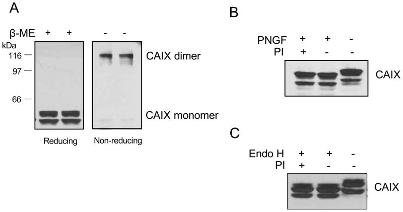
Figure 2. Oligomerization and glycosylation of CAIX in MDA-MB-231 cells.
Panel A: Total membranes were isolated from MDA-MB-231 cells exposed to hypoxia for 16 h. Fifty μg of total membrane protein were separated on an SDS-PAGE gel in the presence or absence of 1% β-mercaptoethanol (β-ME). CAIX expression was detected by western blotting using the M75 monoclonal antibody. Panel B: Total membranes were isolated from MDA-MB-231 cells exposed to hypoxia for 16 h. Fifty μg of protein was digested with 2 μL N-glycosidase F (PNGF) in the presence of absence of protease inhibitor (PI) for 2 hours at 37°C. Panel C: Cell lysates were isolated from MDA-MB-231 cells exposed to hypoxia for 16 h. Fifty g of protein was treated with 2 μL endoglycosidase H (endo H) in the presence or absence of protease inhibitor (PI) for 2 hours at 37°C. CAIX expression was detected by western blotting using the M75 monoclonal antibody. These blots represent duplicate experiments.

