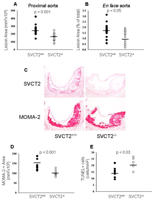
Figure 1.
Atherosclerosis, SVCT2 expression and apoptosis in mouse atherosclerotic lesions. (A, B) Image analysis data obtained from serial sections of aortic sinus (A) and en face aorta (B) of mice reconstituted with apoE−/− FLCs either expressing the SVCT2 (SVCT2+/+, n = 18) or lacking it (SVCT2−/−, n = 19). (C) Immunostaining of sequential random aortic sinus sections for the SVCT2 (top panels) and macrophages (MOMA-2, bottom panels) in apoE−/− mice transplanted with SVCT2+/+ or SVCT2−/− FLCs. (D) Image analysis of MOMA-2 immunostaining from mice transplanted with SVCT2+/+ FLCs or mice transplanted with SVCT2−/− FLCs. (E) Image analysis of TUNEL-positive cells in proximal aortic lesions from mice transplanted with SVCT2+/+ FLCs or from mice transplanted with SVCT2−/− FLCs. In A, B, D and E, bars indicate mean values.

