FIGURE 2.
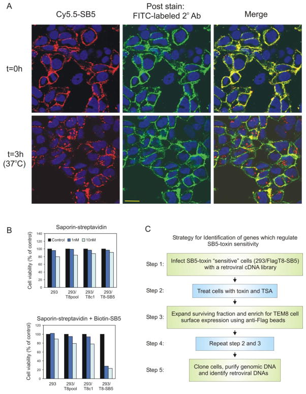
SB5-saporin immunotoxins are internalized and selectively kill 293/T8-SB5 cells. A, Cy5.5-labeled SB5 (red) was taken up into 293/T8-SB5 cells after 3 hours at 37°C. Some of the Cy5.5-SB5 label was detected on the cell surface following the 3 hour incubation, which was confirmed by post-incubation staining with FITC-labeled secondary mAbs (green). The secondary antibody only recognizes primary Cy5.5-SB5 antibody that is present at the cell surface and provides extra sensitivity because of the added layer of amplification. Although some Cy5.5-SB5 antibody was detected at the cell surface, much of the primary antibody had clearly internalized (red, bottom right panel). B, 1 nM or 10 nM of saporin-streptavidin toxin combined with biotin-labeled SB5 selectively killed 293/T8-SB5 cells (bottom panel) compared to saporin-streptavidin alone (top panel). C, Strategy for identification of genes that regulate SB5-toxin sensitivity.

