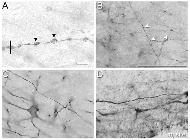
Figure 1.

(A) Representative photomicrograph of dendritic varicosities (DVs) in neurokinin-1 receptor expressing neurons of the rostral ventromedial medulla (RVM) demonstrating the clarity with which DVs could be viewed with a 60× N.A. 1.4 objective and 10× eyepiece objective. The vertical line drawn perpendicular to the dendrite’s trajectory illustrates how the diameter of a DV was determined. Note that the segments between the DVs are constricted. (B) Representative photomicrographs of DVs in neurokinin-1 receptor expressing neurons of the rostral ventromedial medulla 1 hr after microinjection of 100 pmol substance P. (C) Few DVs were observed in neurokinin-1 receptor expressing neurons in the RVM after microinjection of saline. (D) Substance P did not produce DVs in serotonergic neurons in the RVM. Scale bar is 10 μm in panel A and 100 μm in panels B–D. Arrowheads indicate DVs. Where not visible in panels B–D, the microinjection site was within 250 – 500 microns.
