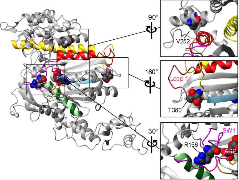
Fig. 3.
Homology model of the crystal structure of the Myo1c motor domain showing the positions of three residues that are associated with hearing disorders when mutated. The regions around each of the three residues are enlarged and rotated for the best view (right). The model was created with Swiss-Model [32] using the solved crystal structure of Dictyotelium MyoE (ADP.vanadate structure) as a template (PDB 1LKX-A). Color-coded structural elements: magenta switch 1 and switch 2, orange P-loop, green SW-2 helix, sky blue β-stand 5, red helix G and loop 1, yellow helix O and HO-linker, dark pink myopathy loop. Bound nucleotide (ADP) and point mutations are shown in space-fill mode

