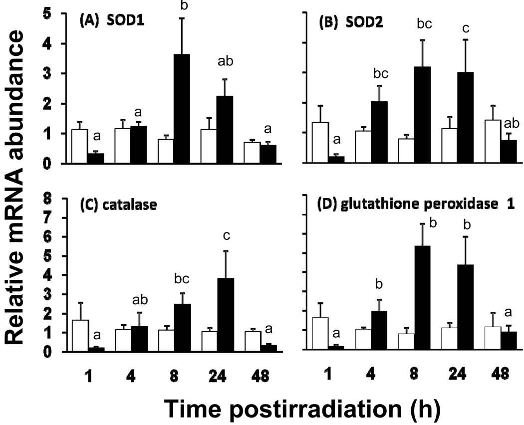
Fig. 3.
Relative mRNA abundance of antioxidant enzymes in isolated jejunum of unirradiated (open bars) and irradiated (8.5 Gy, filled bars) mice. (A) SOD1, (B) SOD2, (C) catalase, and (D) glutathione peroxidase 1 as measured by real time PCR. mRNA was collected from the mucosa of the proximal small intestine of mice fed with control diet and sacrificed 1, 4, 8, 24 and 48 h post-irradiation. Results are means ± SE (n = 6). Filled bars with different superscripts are significantly different (P < 0.05 by one-way ANOVA). Thus, there is a marked decrease in levels of mRNA at 1irradiation, followed by a dramatic increase, and then a decline to levels similar to unirradiated mice. Open bars are similar throughout, indicating that expression in unirradiated mice did not change over time.

