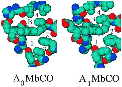
Figure 2.
X-ray-determined structure of left Mb at pH 5 (Protein DataBank file 1spe, from ref. 38), and right Mb at pH 7 (Protein Data Bank file 1a6g, from Ref. 23). His-64 (Upper Left) has moved outside the heme pocket in A0, and smaller changes are visible throughout the protein. The B-site is labeled B, and the xenon cavities 1, 2, 3, and 4 are labeled with numbers.

