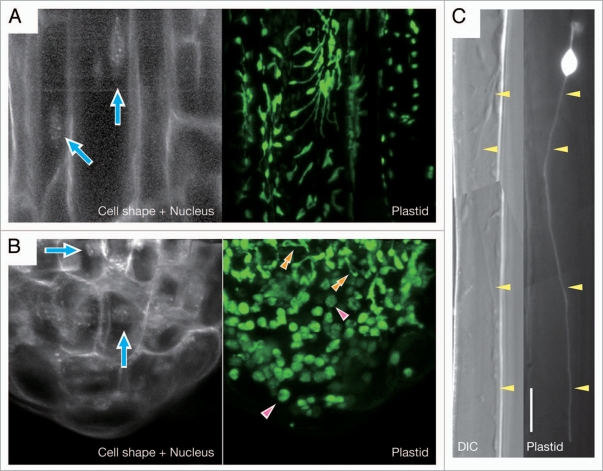
Figure 1.
Morphology of non-green plastids in Arabidopsis thaliana roots. Plastids in the epidermal cells of main roots (A and C) and the root tip, including root cap and meristem (B), were visualized by plastid-targeted green fluorescence protein, pt-sGFP(S65T).15 For each panel, GFP images are shown at the right. For (A and B), 4′,6-diamidino-2-phenylindole (DAPI)-stained fluorescent image of the same field of view as the GFP image is at the left to show the positions of cell nuclei (indicated by arrows) and the cell outlines. In (B), single arrowheads and double arrowheads indicate mature amyloplasts in the columella cells and transitional plastids that are differentiating into amyloplasts from proplastids, respectively. For (C), the differential interference contrast (DIC) image of the same field of view as the GFP image is at the left to show the presence of an exceptionally long stromule (indicated by arrowheads), which reaches a length of about 96.8 µm including the length of the plastid main body. Bar = 10 µm.

