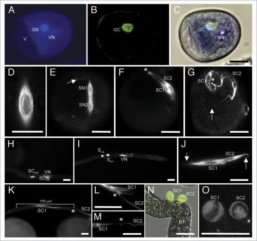
Figure 1.
Establishment and properties of the male germline in maize. Germline cells are labeled with α-tubulin-YFP. (A–D) Microspore at late bicellular stage: (A) the vegetative cell still contains a vacuole (V) and the generative cell nucleus (GN) already underwent DNA-syntheses as indicated by its bright signals compared with the vegetative nucleus (VN) after DAPI staining. (B) α-tubulin-YFP expression is restricted to the generative cell (GC). (C) Merged image of (A and B). (D) α-tubulin-YFP forms a cage around the GC nucleus during Pollen Mitosis II . (E) Transition towards tricellular stage: microtubuli bundles have been formed around and between sperm nuclei (SN1/2). Note that first microtubular tail-like extensions are visible (arrow). (F) At early tricellular stage, twin sperm cells (SC1/2) are arranged in parallel. A microtubuli knot (asterisk) becomes visible at half distance between sperm nuclei. (G) Late and mature tricellular pollen stage showing twin sperm cells with long microtubular extensions (arrow), a microtubuli knot (asterisk) connecting both cells and the germination pore (P). (H and I) DAPI staining to show that sperm cells and vegetative nucleus travel as male germ unit (MGU). Note that initially the nucleus of the leading sperm cell (SCVN) seems closely associated with the vegetative nucleus (VN). (I and J) At later stages leading and trailing sperm cells (SCVN and SCUA) are hardly distinguishable as they change positions inside the growing pollen tube. Arrows mark tail-like microtubuli extensions. (K) Stretched sperm cells measure up to 155 µm in length. An asterisk labels the position of a microtubuli knot between both sperm cells. (L and M) Examples of enlarged microtubuli knots. (N) A manipulated pollen tube (PT) releases twin sperm cells that become spherical within seconds. (O) Spherical sperm cells have completely lost their microtubular structure. Dark areas inside sperm cells are sperm nuclei. Scale bars: 20 µm.

