FIGURE 2.
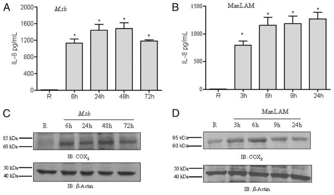
M. tuberculosis H37Rv infection or ManLAM stimulation enhances IL-8 production and COX2 expression in human macrophages. The cell culture media and cell lysates of MDMs incubated with M. tuberculosis H37Rv or stimulated with ManLAM were analyzed for IL-8 production by ELISA and COX2 expression by Western blot, respectively. The time points were the same as in Fig. 1. The media of uninfected macrophages showed undetectable levels of IL-8. In contrast, the media of M. tuberculosis-infected cells (A) or ManLAM-stimulated cells (B) showed significantly increased levels of IL-8. Shown are cumulative results of three independent experiments performed in triplicate in A and five independent experiments performed in triplicate in B (mean ± SEM). *p < 0.0001. Cell lysates from M. tuberculosis-infected cells showed increased COX2 expression relative to uninfected cells (C). MDMs stimulated with ManLAM showed increased expression of COX2; the expression peaked at 6 h and was sustained until 9 h (D). The Western blots shown are representative of three independent experiments. Immunostaining for β-actin was used as a loading control.

