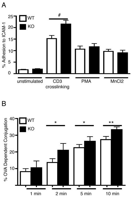
Figure 3. PIPKIγ90 −/− CD4+ T cells exhibit increased adhesion to ICAM-1 and increased conjugation to APC.
a) Anti-CD3/28 bead activated CD4+ T cells were fluorescently labeled and adhered to ICAM-1 coated plates untreated or following stimulation with anti-CD3 crosslinking, PMA or MnCl2. Following washing step, the percentage of cells adherent to the plate was determined. Results are averages from 3 independent experiments +/− SEM. #=p<0.01 compared to wild type anti-CD3 treated. B) OVA peptide expanded CD4+ T cells were calcein labeled and incubated with PKH-26 labeled LB27.4 cells +/− 2.5 ug/ml OVA peptide for the indicated times. Non-specific conjugates were dissociated by vortexing and the percentage of OVA-dependent conjugation determined by flow cytometry. Results are averages from 5 independent experiments +/− SEM *=p<0.05 **=p<0.0001 compared to wild type.

