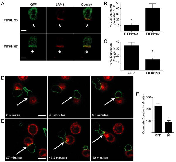
Figure 5. Overexpression of PIPKIγ90 impairs T cell conjugation and decreases duration of T cell-APC contact.
A) D10 T cells overexpressing GFP-PIPKIγ90 or PIPKIγ87 were fixed and stained with anti-LFA-1 antibodies following conjugation to conalbumin loaded CH12 cells (represented by star). Scale bar represents 5um. Images are representative of over 60 conjugates observed in 4 independent experiments. B) Quantification of GFP polarization in T cells overexpressing GFP-PIPKIγ90 or PIPKIγ87. Percentage of conjugates with polarized GFP was determined by blinded observation of over 60 conjugates from 4 independent experiments. *=P<0.05 C) D10 T cells overexpressing GFP or PIPKIγ90 were allowed to interact with PKH-26 labeled CH12 cells +/− conalbumin for 2 minutes prior to dissociation of non-specific conjugates. Percentage of antigen dependent conjugates was determined by flow cytometry. Data are mean +/− SEM from 3 independent experiments. *=P<0.05. D and E) Time-lapse microscopy of GFP-PIPKIγ90 overexpressing cells with PKH26 labeled APC. Scale bar represents 10 um. D) PIPKIγ90 is redistributed from the uropod following T cell-APC contact. E) PIPKIγ90 reestablishes uropod localization following conjugation. Images from C and D are representative of over 10 conjugation events observed in 4 independent experiments. F) Quantification of T-cell-APC duration in T cells overexpressing GFP or GFP PIPKIγ90. Data represent means +/− SEM from 4 independent experiments of at least 10 conjugation events. *=P<0.01.

