
Figure 6. PIPKIγ90 −/− CD4+ T cells exhibit increased proliferation in response to CD3/CD28 coated beads and PMA/Ionomycin.
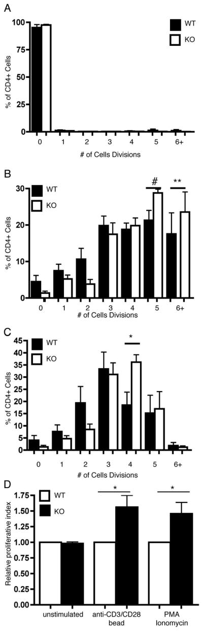
OVA peptide expanded CD4+ cells were stained with 0.25 uM CFSE and left unstimulated (a) or stimulated with anti-CD3/CD28 coated beads (B) or PMA/Ionomycin (c). CFSE dilution was measured 72 hours following stimulation. The % of cells in each cell division and proliferative index were determined by ModFIT analysis. D) Normalized proliferative index from 5 independent experiments. Data represent averages +/− SEM from 5 independent experiments *=p<0.05, **=p<0.01. #=P=0.065