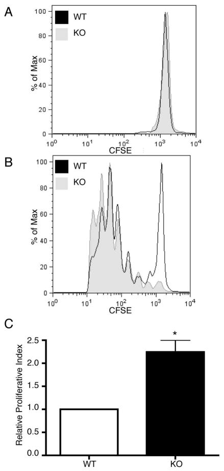
Figure 7. PIPKIγ90 −/− CD4+ T cells exhibit increased proliferation in vivo.
Naïve CD4+ T cells from wild type and PIPKIγ90 −/− mice were labeled with 2.5 uM CFSE and injected intravenously into age/sex matched recipients. 18 hours later, mice were given an IP injection of 25 ug LPS (A) or 25 ug LPS and 50 ug ovalbumin(B). 72 hours later, CFSE dilution was measured in CD4+ T cells by flow cytometry and proliferative index (C) determined by ModFit analysis. Data are representative of 2 independent experiments involving 3 wild type and knockout mice per condition.

