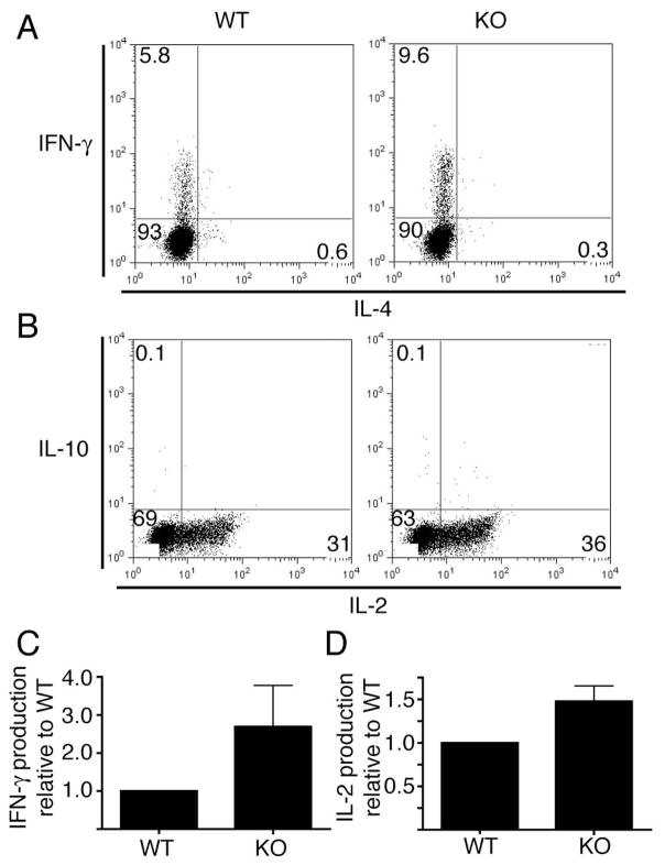
Figure 8. PIPKIγ90 −/− CD4+ T cells have increased Th1 cytokine production in vitro.
A and B) OVA peptide expanded CD4+ cells were restimulated on day 7–10 post isolation with plate bound anti-CD3 and soluble CD28 in the presence of brefeldin A. Four hours later, cells were stained with anti-CD4, fixed, permeabilized, stained with antibodies to IFN-γ and IL-4 (A) or IL-2 and IL-10 (B), and analyzed by flow cytometry. Results are representative of 3 independent experiments with cells from 3 wild type and knockout mice. C and D) Normalized IFN-γ (C) and IL-2 (D) producing CD4+ cells +/− SEM from three independent experiments.

