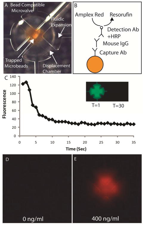
Figure 6.
(A) Trapping microvalve loaded with approximately 120 ng of magnetic microbeads coated with antimouse IgG. The bead-compatible digital microfluidic Automaton uses microvalves with larger fluidic expansions to prevent the beads from becoming trapped as they are transferred through the array. (B) Schematic of the immune complex formed in the immunoassay for mouse IgG. (C) Fluorescence profile obtained while rinsing 10 uM fluorescein dye from the trapping microvalve. The same program was used to remove unbound analyte and detection antibody after their corresponding incubations. Epifluorescence images of trapping valves after performing the assay with a negative control (D) and 400 ng/ml mouse IgG sample (E).

