Fig. 1.
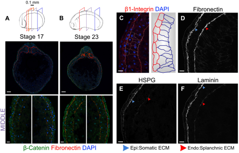
Bilayered LPM is LR symmetrical from tailbud-tadpole stages with splanchnic-somatic structural differences beginning at stage 23. (A,B) Diagrams indicate stage/length and sectional planes. Analysis every 0.1 mm (dashed red frame) was between anterior-posterior LPM extremes indicated by red/blue frames. Representative mid-embryo sections (purple frame) are shown. (A) Stage 17 (10×, 40×), left and right LPM each comprising two layers. β-Catenin (green); DAPI (blue). Fibronectin (red) flanks epidermal/endodermal faces of left and right LPM. (B) Stage 23: maintenance of bilayered left and right LPM. (C) Left/right LPMs are structurally similar during these stages, but somatic/splanchnic layers become distinct, symmetrically, from stage 23; somatic cells are more squamous, splanchnic are more columnar. (D-F) Somatic and splanchnic LPM show different basal lamina compositions. Somatic: strong fibronectin, HSPG and Laminin signal; splanchnic: much weaker HSPG/Laminin signal, especially laterally. Scale bars: 100 μm, in top images A,B; 20 μm in bottom images A,B; 20 μm in C-F.

