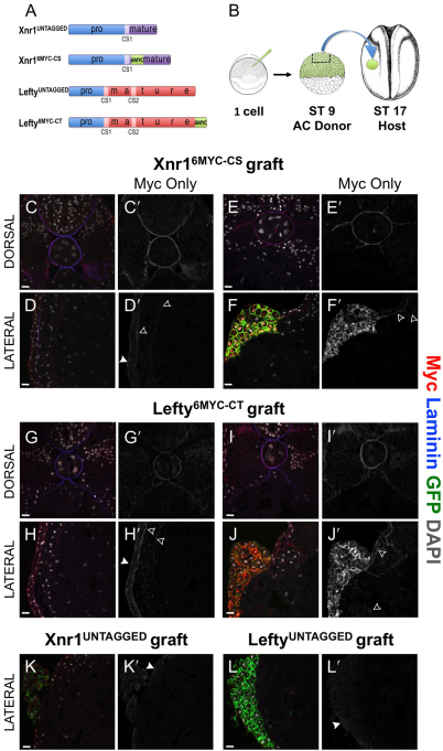
Fig. 3.
Xnr16MYC-CS and Lefty6MYC-CT move substantially from AC grafts. (A) Xnr1 and Lefty constructs: blue box, pro-domain; CS1/CS2, cleavage sites liberating mature ligands; 6MYC tag was inserted just downstream of CS1 (Xnr1) or C-terminally (Lefty). (B) AC-grafting schematic. (C-L′) Transverse cryosections were used to detect Myc (red; grayscale in C′-L′), laminin (blue) and nuclei (DAPI, white); dorsal panels focus axially/paraxially, lateral panels on LPM. Membrane-bound GFP (mGFP, green) marks engrafted cells; 2.5 μm optical sections. Open arrowheads, Myc; closed arrowheads, nonspecific epidermal haze. (C-F′) Xnr16MYC-CS, (G-J′) Lefty6MYC-CT, (K,K′) Xnr1UNTAGGED and (L,L′) LeftyUNTAGGED. (C,C′,D,D′) Representative section ∼110 μm anterior of graft margin; Xnr16MYC-CS signal in basal lamina surrounding notochord/neural tube. Dorsal and left LPM Xnr16MYC-CS signal colocalized with laminin. (E,E′,F,F′) Representative images, dorsal and lateral Xnr16MYC-CS signal within/near graft. Note absence of endoderm signal. (G,G′,H,H′) Lefty6MYC-CT signal colocalized with laminin in dorsal and lateral views, ∼340 μm anterior of graft. (I,I′,J,J′) Dorsal and lateral images of Lefty6MYC-CT signal. Note Lefty6MYC-CT signal is within endoderm, not colocalized with laminin. (K-L′) AC grafts with Xnr1UNTAGGED or LeftyUNTAGGED reveal artefactual hazy epidermal signal (closed arrowheads). Scale bars: 25 μm.

