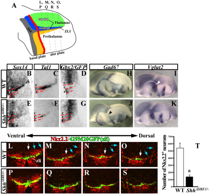
Fig. 5.
Maturation of rostral thalamic neurons is dependent on the ventral midline source of Shh. (A) Schematic representation of a sagittal view through the mouse diencephalon (see Fig. 4A for details) showing the plane of sections in L-S. (B-G) In situ hybridization of coronal sections through wild-type (WT) and ShhΔSBE1/– mutants at E12.5 stained for markers of postmitotic neurons derived from pTH-R (Sox14 and Tal1) and pTH-C (Gbx2). The red dashed lines mark the borders between caudal (c), rostral (r) and zli (z) regions of the thalamus. The Shh-GFP reporter line was used to mark the zli in D and G. (H-K) Gad67 (pTH-R neurons) and Vglut2 (pTH-C neurons) expression in WT and ShhΔSBE1/– embryos. The black arrowhead in J indicates the reduction in Gad67 expression. The gap in Vglut2 expression (white arrowhead in I) is filled in K. (L-S) Nkx2.2 (pTH-R) and GFP (zli) immunostaining on coronal sections through WT and ShhΔSBE1/ embryos at E12.5. Nkx2.2 expression was reduced in both pTH-R progenitors (white arrows) and postmitotic neurons (blue arrows). (T) The number of Nkx2.2+ neurons was significantly reduced in ShhΔSBE1/– embryos (*P<0.005). Data are expressed as mean ± s.e.m. pTH-C, caudal thalamic progenitors; pTH-R, rostral thalamic progenitors.

