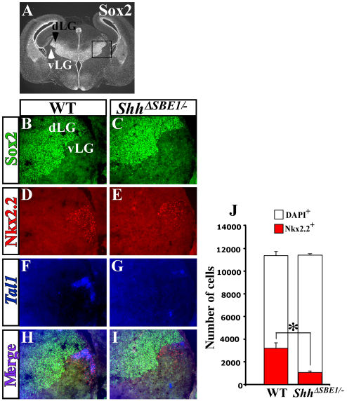
Fig. 6.
vLG neurons are dependent on ventral midline Shh for specific aspects of their differentiation. (A) Sox2 immunostaining shows a sharp boundary between the dorsal lateral geniculate nucleus (dLG, black arrowhead) and ventral lateral geniculate nucleus (vLG, white arrowhead) in a wild-type mouse embryo at E16.5. (B-I) Triple labeling for Sox2, Nkx2.2 and Tal1 on coronal sections through wild-type (WT) and ShhΔSBE1/– embryos at E16.5. Merge of triple images shows overlap of the expression of Nkx2.2 and Tal1 in vLG neurons from wild-type (H) but not ShhΔSBE1/– embryos (I). (J) Quantification of Nkx2.2 (red) and DAPI (white) positive cells in the vLG of WT and ShhΔSBE1/– embryos. A significant reduction in the number of Nkx2.2+ neurons was observed in ShhΔSBE1/– embryos (*P<0.005) whereas the total number of vLG neurons was unaffected. Data are expressed as mean ± s.e.m.

