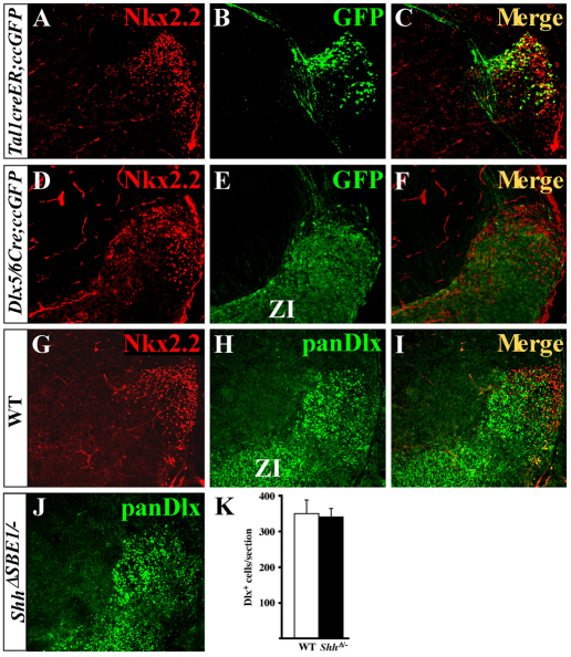
Fig. 7.
Dual origin of vLG neurons in mouse. (A-C) Tal1creER;ccEGFP embryos were exposed to tamoxifen at E11.5, harvested at E16.5 and stained for Nkx2.2 and GFP. (D-F) Dlx5/6Cre;ccEGFP embryos were immunostained for Nkx2.2 and GFP at E16.5. (G-I) Nkx2.2 and panDlx antibodies mark distinct neurons in the vLG at E16.5. (J,K) The number of Dlx+ cells in the vLG in ShhΔSBE1/– embryos (J,K) is comparable with that observed in wild type (H). Data are expressed as mean ± s.e.m. ZI, zona incerta.

