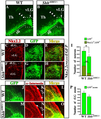
Fig. 8.
Tracing the fate of pTH-R neurons in wild-type and ShhΔSBE1/– embryos. (A,B) Nkx2.2cre; ccEGFP embryos at E16.5 stained for GFP on coronal sections through wild-type (WT; A) and ShhΔSBE1/– (B) embryos. GFP-positive cell bodies (arrow) and axons (arrowheads) are shown. Boxed area indicates magnified region in C-H. (C-H) Colocalization of Nkx2.2 and GFP-expressing neurons in the vLG of WT (C-E) and ShhΔSBE1/– (F-H) embryos. (I) Quantification of data from E and H. Data are expressed as mean ± s.e.m. *P<0.05, **P<0.005. (J-O) Colocalization of GFP and α-internexin (intermediate filament marker) on caudally projecting vLG axons from WT (J-L) and ShhΔSBE1/– (M-O) embryos. The majority of GFP+ neurons and caudally projecting axons are present in the vLG of ShhΔSBE1/– mutants compared with wild-type littermates despite the reduction in Nkx2.2 expression. Arrows in K and N point to the optic tract (OT). (P) Quantification of data from J and M. Data are expressed as mean ± s.e.m. P<0.05. dLG, dorsolateral geniculate; Th, thalamus; vLG, ventrolateral geniculate nucleus; zli, zona limitans intrathalamica.

