Fig. 1.
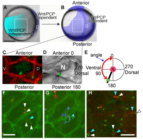
Embryonic regions examined, methods for MTOC position measurement and some functions of centrin-labeled MTOCs. (A) In a midgastrulation embryo, Wnt/PCP signaling is required in cells of the dorsal midline (D) for convergence and extension, but not in lateral regions. The regions within the dotted white line were examined for MTOC angle. Scale bar: 200 μm. (B) Late gastrulation embryo. Lateral mesodermal cells flank the notochord and require Wnt/PCP signaling to migrate efficiently. (C) Membrane (red) and centrosome (green) labeling. Cells were oriented so that anterior of the embryo was oriented towards the top of the frame during measurement. Dorsal (D) and ventral (V) in the embryo are indicated. (D) Nucleus is visible in DIC optics with a green dot denoting an overlain MTOC. Dotted black line indicates orientation of MTOC relative to the nucleus. Distance between MTOC and nucleus was not measured. (E) Illustration of how MTOC position was measured relative to the nucleus N and the embryonic body axes. Angle measured is indicated by the red line. (F) Centrosomes (Xenopus centrin-Cherry, white arrowhead) in projected z-stack have a membrane connection to the cell membrane. (CAAX-EGFP, blue arrowhead). (G) Centrosomes move in cells. Paths followed for 10 minutes start at the square. (H) Short cilia (red, acetylated tubulin, white arrowhead) colocalize with some centrosomes (eGFP-Xcentrin, blue arrowheads). Scale bars: 10 μm.

