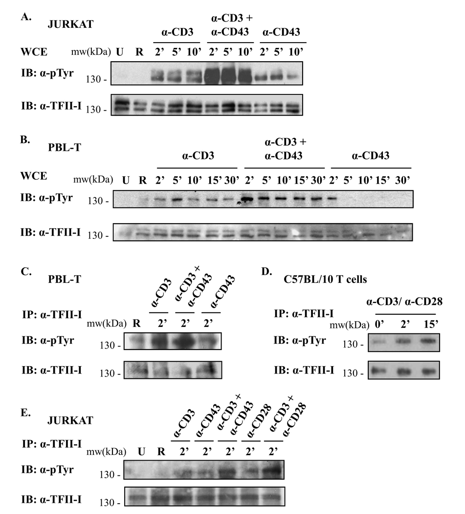
Figure 2. Simultaneous crosslinking of two receptors results in stronger TFII-I phosphorylation than crosslinking receptors singly.
(A) Jurkat cells and (B) human peripheral blood lymphocytes (PBL-Ts) were serum-starved and stimulated with either anti-CD3 + RaMIg, anti-CD3 + anti-CD43 + RaMIg, anti-CD43 + RaMIg Abs or RaMIg alone (R lane) for the times specified. Phosphotyrosine levels of TFII-I in whole cell extracts (WCE) were detected by immunoblotting using an anti-p-Tyr Ab (upper panels). Blots were stripped and re-probed with anti-TFII-I Ab to reveal TFII-I protein levels (lower panels). U lane: unstimulated cells. (C) PBLTs, (D) splenic T cells from C57BL/10 mice and (E) Jurkat cells were serum-starved and stimulated either as described in (A, B) but for 2’ or with anti-CD3 + anti-CD28 Abs for the times indicated. WCE were then subjected to IP with anti-TFII-I Abs. Phosphotyrosine levels of TFII-I were detected by immunoblotting (IB) using an anti-pTyr Ab. Blots were stripped and re-probed to reveal TFII-I protein levels using anti-TFII-I Ab. Data are representative of at least two independent experiments for (A –E).

