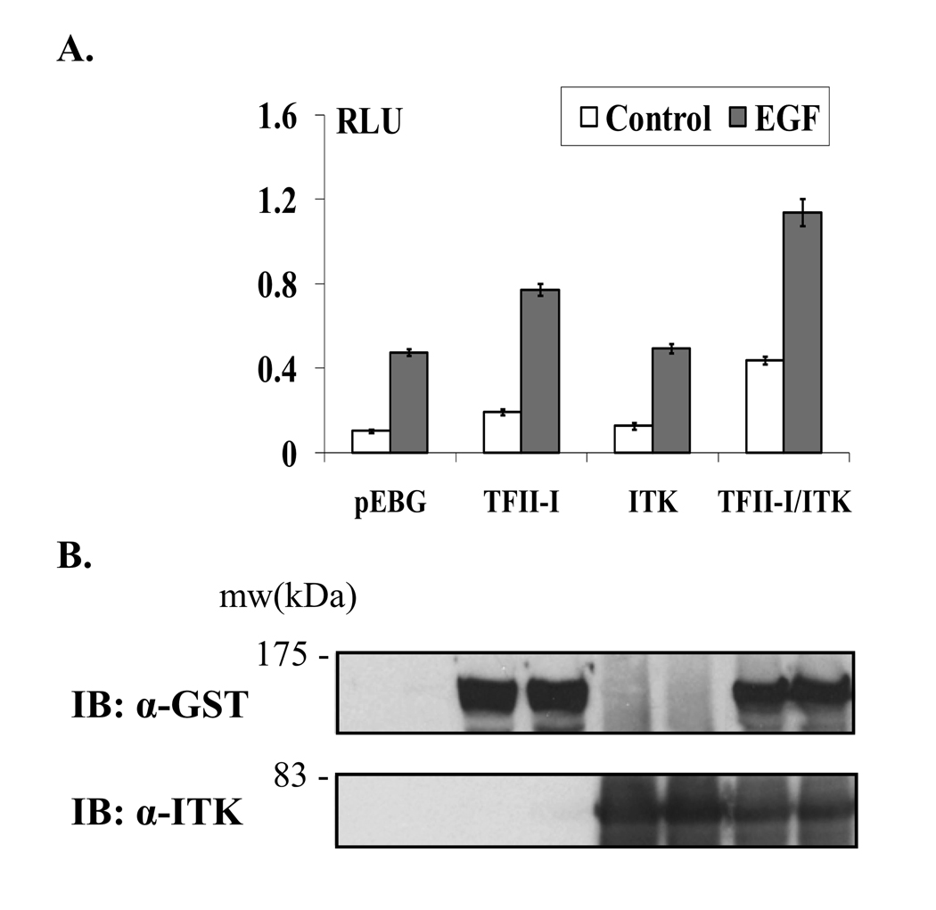
Figure 6. Itk potentiates the TFII-I-driven transcriptional activity of the c-fos promoter under basal and growth factor-stimulating conditions.
(A) Wild type TFII-I-Δ-GST was expressed in the presence or absence of wild type ITK-Myc in COS-7 cells, along with a c-fos promoter luciferase construct and the levels of transcription measured by a luciferase reporter assay. The pEBG empty vector was used as a negative control in the assay. Transfected cells were either stimulated with EGF for 4 h (EGF columns) or remained untreated in serum-free medium (control columns). RLU: relative luciferase units. Transfections were performed in triplicate and the results presented as mean ± SD. (B) Western blot analysis of equivalent volumes (50µl) of the COS-7 whole cell lysates used in the luciferase reporter assay. TFII-I-Δ-GST protein levels were visualized using anti-GST Ab (top panel). The membrane was stripped and re-probed using anti-Itk Ab to monitor Itk-Myc protein levels (bottom panel). The results are representative of at least two experiments for (A) and (B).

