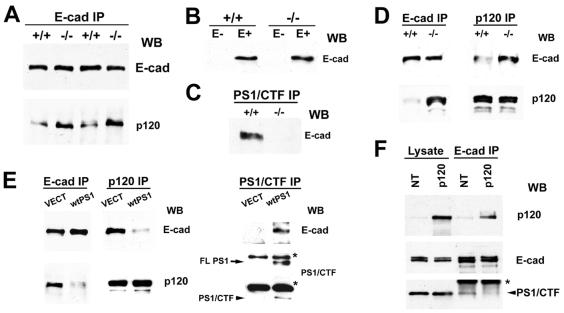
Figure 3.
PS1 inhibits p120 binding to E-cadherin. (A) E-cadherin IPs (E-cad IP) in TNE plus 1% Triton X-100 from either PS1−/− mouse embryos (−/−) or their PS1+/+ littermates (+/+) were probed with anti-E-cadherin (E-cad) or anti-p120 (p120) antibodies. (B) Extracts (1% SDS) of confluent nontransfected (E−) or E-cadherin-transfected (E+) PS1+/+ (+/+) and PS1−/− (−/−) mouse fibroblasts were probed with anti-E-cadherin antibody. (C) Extracts (1% Triton X-100) of confluent E-cadherin-expressing PS1+/+ (+/+) or PS1−/− (−/−) fibroblasts were immunoprecipitated with antibody 33B10 (PS1/CTF IP), and the resulting IPs were probed with anti-E-cadherin (E-cad) antibody. (D) Extracts (1% Triton X-100) of E-cadherin-transfected PS1 +/+ (+/+) or PS1−/− (−/−) fibroblasts were immunoprecipitated with anti-E-cadherin (E-cad IP) or anti-p120 (p120 IP) antibodies, and the resulting IPs were probed with anti-E-cadherin (E-cad) or anti-p120 (p120) antibodies. (E) E-cadherin-transfected PS1−/− fibroblasts were transiently transfected either with vector (VECT) or with WT PS1 (wtPS1), and Triton X-100 extracts were immunoprecipitated with anti-E-cadherin (E-cad IP), anti-p120 (p120 IP), or anti-PS1/CTF (PS1/CTF IP) antibodies. The resulting IPs were probed with anti-E-cadherin (E-cad), anti-p120 (p120), or anti-PS1/CTF antibodies. FLPS1, full-length PS1. The asterisks indicate IgGs. (F) Extracts (1% Triton X-100) of untransfected (NT) EL cells or EL cells transiently transfected with p120 (p120) were treated with anti-E-cadherin antibodies (E-cad IP), and the resulting IPs were probed with antibodies against the proteins indicated on the right. The asterisk indicates IgGs.

