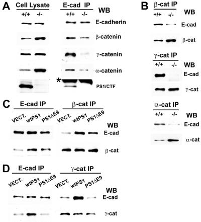
Figure 5.
PS1 stabilizes the cadherin/catenin complex. (A) Extracts of confluent E-cadherin-transfected PS1+/+ and PS1−/− fibroblasts in 1% Triton X-100 were treated with anti-E-cadherin antibody, and the resulting IPs (E-cad IP, Right), along with total cell lysate in 1% SDS (Left), were probed on WBs with antibodies against the proteins indicated at the right of the figure. The asterisk denotes low-molecular-weight IgGs. (B) Anti-β-catenin, anti-γ-catenin, or anti-α-catenin IPs (Top, Middle, and Bottom, respectively) prepared from E-cadherin-expressing PS1+/+ and PS1−/− fibroblasts were probed with anti-E-cadherin (E-cad), anti-β-catenin (β-cat), anti-γ-catenin (γ-cat), or anti-α-catenin (α-cat) antibodies. (C and D) Extracts (1% Triton X-100) of E-cadherin-expressing PS1−/− fibroblasts transiently transfected with vector (VECT), WT PS1 (wtPS1), or PS1 FAD mutant ΔE9 (PS1ΔE9) were treated with anti-E-cadherin (E-cad IP), anti-β-catenin (β-cat IP) (C), or anti-γ-catenin (γ-cat IP) (D) antibodies, and the resulting IPs were probed with antibodies against the proteins indicated at the right of the figure. E-cad, E-cadherin; β-cat, β-catenin; γ-cat, γ-catenin.

