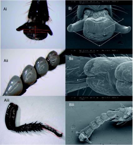
Figure 2.
A) Light (LM) and B) scanning electron (SEM) images of i) Pre-tarsal organ (magnification 40× LM, S3.1× SEM) ii) Euplantula (mag. 30× LM, 88.0× SEM) iii) Entire foot of Gromphadohina portentosa (mag. 6.3× LM, 15.9× SEM). Red lines indicate measurements taken, white circle demonstrates area where “finger-like projections” were found. Labels; a = Arolium, p = Pretarsal claws, and e = Euplantula. High quality figures are available online.

