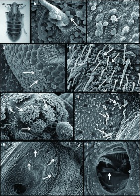
Figure 1.
Scanning electron micrographs: (a) Photo of adult females of Peregrinus maidis infected with Beauveria bassiana CEP189 and Metarhizium anisopliae CEP 160, Bar: 0.7mm (b) B. bassiana conidia over a hair socket on the antennae (arrow), Bar: 2 ìéôé. (c) B. bassiana germ tube penetrating through a pore of the wax glands (arrow), Bar: 2 µm. (d) B. bassiana conidia between the ommatidia of the compound eye (arrows), Bar: 20 µm. (e) B. bassiana conidia in the second antennal segment (arrows), Bar: 10 µm. (f) B. bassiana conidia between the hairs of the antennal sensory pits (arrows), Bar: 20 µm. (g) M. anisopliae conidia near to the pores of the wax glands (arrows), Bar: 11.7 µm. (h) M. anisopliae conidia near the spiracle (arrows), Bar: 14 µm. (i) M. anisopliae conidium enclosed within the spiracle (arrow), Bar: 4 µm. High quality figures are available online.

